More On Your Nerves

neurodynamics1.jpg

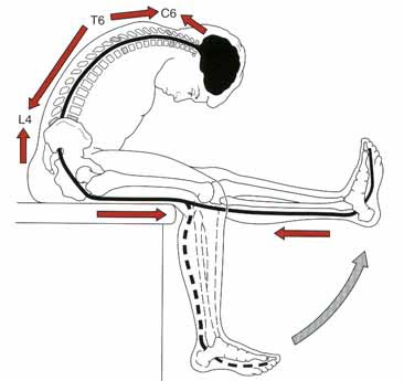
In part one of this series I highlighted the connection between the Nervous System and the Immune System.   Previously viewed as two separate entities, these can now confidently be viewed as one interconnected system.  However it is this exact relationship that influences the physical capacity of our nerves and driving the neural symptoms often experienced in the arms and legs.  In part two, we will discuss neurodynamic testing as a tool to evaluate these physical capacities and how they may be restricting our movement patterns.

It may be surprising, but our neural structures are the most resilient human tissue we have.  Think about how adaptive they must be in real time. Every step we take, door we open, or Saturday night dance move we bust is only achievable because our nerves are able to efficiently adapt.  If our nerves did not glide, lengthen, and shift none of this would be possible without eliciting neural tension. A happy nerve demands, blood flow, space, and movement to function optimally and the restoration of these is fundamental to treatment.  

But how do you know which is needed?  Insert neurodynamic testing.

neurodynamics2.jpeg

To be effective with neurodynamic testing there has to be an understanding of the science between normal neural mechanics and neural physiology.   In other words, neurodynamics assess and treat the physical health of the nervous system. When pathology exists in either the mechanics or physiology of a nerve it can disrupt the conductivity of the nerve making the communication between the nerves, brain, and muscles inefficient.  This can be difficult to treat effectively, if not identified correctly. However the use of neurodynamic testing intentionally positions and loads a nerve to help communicate where the dysfunction is stemming from.  This testing thereby identifies the capacity of a nerve to slide, glide, angulate, strain or compress efficiently.  

* Fun fact:  A peripheral nerve can lengthen approximately 12-20% to accommodate normal movement, while the spinal cord can elongate nearly an inch!  So, those tight hamstrings may not be due to muscle tightness, if you have symptoms into the legs, it could be a restriction within the nervous system.

Neurodynamic tests are thus designed to bias neural elements more so than the surrounding interfaces. This intentional loading of a specific nerve root, trunk, and localized region of the spinal canal, and/or sympathetic nervous system allows us to grasp a deeper understanding of where the symptoms are being generated from to treat the cause rather than the symptom.   

Here is a video from David Butler to help further explain how our nerves move!  Check it out.

Stay Well, Stay Strong

Keaton

Practical Stress Management: Part 2

Screenshot 2018-05-10 at 7.59.30 AM.png

If there is one aspect of life that is often undervalued and under-appreciated it is sleep.  Our ability to sleep is essential to allow a full resolution of pain and restoration of our movement.  Our ability to sleep can be correlated to how our nervous system is being activated. We have 2 parts of our nervous system:  sympathetic and parasympathetic. You see, when we are experiencing pain our nervous system becomes sensitized and preferential to the sympathetic nervous system.  Now when this is the case you will see a rise in heart rate, blood pressure, respiration rate, and even anxiety. Sound familiar on those sleepless nights? You can loosely think of this activation of the sympathetic nervous system as if you drank a large cup of coffee or an energy drink right before bed.  Most people would not choose to partake in such behavior because they would be up all night. This is the exact case for many of those who have had or are currently in a pain-cycle however it isn’t perceived as such. People who are experiencing sleep disturbances or find it difficult to become comfortable at night, please realize that the solution is on the other side of the nervous system:  parasympathetic. There are numerous things we can control or change in the activities leading up to bed as well as with the sleep environment that will allow the parasympathetic nervous system to be tapped into.

According to the National Sleep Foundation, sleep hygiene is considered to be a variety of different practices and habits that are necessary to have good nighttime sleep quality and full daytime alertness.  Sounds great, but what does this have to do with resolving pain and improving movement? Well, consider sleep as the foundation of your health and stress management. By purposefully improving sleep hygiene you are taking a step in regaining control of your life.  Here’s how to do it:

1. Be regular and consistent with the time you get up.  

That’s right when you get up opposed to when you go to sleep.  The reason is that this will allow you to improve your sleep efficiency, the ratio of time in bed asleep opposed to in bed and awake.  The first couple of days may be rough and you may feel fatigue during the day, but this should be reflected on the back end of the day because you will feel tired sooner and be able to determine your ideal bedtime.  I guess you can say we are trying to reverse engineer your bed time.

2. Don’t oversleep.  

Many people live by the idea that they can go on little to no sleep during the week and make up for it on the weekend.  This is not the case. You cannot make up for your accumulated weekly sleep debt by sleeping in on the weekends or days off.  What you are actually doing is setting yourself up for increased grogginess and fatigue throughout the day, which in turn could increase tension and amplify your aches and pains.  

3. Create a positive psychological association with the bedroom.  

The purpose of a bedroom is two-fold:  sleep and sex. If you currently reading, watching television, drinking a cup of tea, etc. before bed, work to try to move these activities to somewhere outside of the bedroom and prior to being ready for sleep.  To perform these activities it requires an increase in alertness and attention, which are both associated with wakefulness and thus an increase in sympathetic activity.

4. Substance avoidance:   alcohol, nicotine, or caffeine.  

Alcohol is often regarded as the ideal nightcap beverage given that it is a depressant.  This is true that it is a depressant, but what is often missed is that alcohol causes us to fall asleep faster than we normally would thus miss out on a portion of our REM (radpid eye movement) cycle.  This is important because disruption of the REM cycle can alter the entire sleep-wake cycle and associated hormonal releases that help you both function and recover when injured.

With regards to nicotine and caffeine, they are both stimulants and will make it difficult to fall asleep.  Now if your thinking I drink a cup of coffee before bed every night and don’t have any changes in my ability to sleep.  I am here to tell you that this is likely due to you already being sympathetically driven and in need of a shift into more parasympathetic activity.  Take the caffeine away and see if you notice a difference.

5. Can’t sleep, GET UP.  

If you lay in bed unable to sleep for more than 20-30 minutes get up and perform some form of relaxation:  meditation, breathing exercises, etc. Once you feel tired again return to your bedroom and go to sleep. The goal here is to keep the positive association with your sleeping environment.  

6. Get rid of all your thoughts and worries.  

A lot of people ponder their to do list for the next day or revisit a stressful part of work day that happened this only achieves a rise in heart rate and blood pressure.  Take the 3-5 minutes before going to bed to let it all out in a journal so you can clear your mind before you sleep.

7. Create a sleeping environment.

Try to implement 1 or more of these sleep environment changes to optimizes your quality of sleep once you fall asleep.  

Screenshot 2018-05-10 at 7.39.11 AM.png

I hope these help you understand and appreciate the role of sleep in our ability to not only manage stress in our lives, but how it can facilitate an improvement in function.  In the next post we will visit the ever controversial topic of nutrition.

Stay Well, Stay Strong, 
Keaton

References:

  1. Chouchou, F. and Desseilles, M. (2017). Heart rate variability: a tool to explore the sleeping brain?.

  2. H. Craig Heller, P.  (2017.) https://www.thegreatcourses.com/.

  3. Hartman, B.  (2017). All Gain, No Pain.

  4. sleepfoundation.org.  (2017).  Sleep Hygiene.  [online] Available at:  https://sleepfoundation.org/sleep-topics/sleep-hygiene [Accessed 21 Oct. 2017].


Practical Stress Management: Part 1

stressmanagement.jpeg

Recently we have highlighted the impact that stress can have on our breathing pattern as well as our movement, but how do we effectively manage stress?  This is the first in a 4 part series emphasizing specific behaviors that can optimize stress management. In this post we will discuss the importance of establishing a morning ritual to help set up a day for success.  This will be followed by sleep optimization, nutritional support, and concluded with the benefits of consistent exercise.

Let’s jump into learning the importance of a morning ritual.  This will be the easiest of the four pillars to implement into life immediately.  

What is a morning ritual?

First, I believe that we have to define, ritual.  A ritual, typically associated with religious beliefs, can be viewed secularly as an order of actions arising from convention or habit to support a goal.(1)  It is likely that most, if not all, of us already have some form of a ritual each morning. But is it setting a foundation for success? The key component of a ritual should be the freedom of thought.(1)  We should not have to think about what our next action will be. This is wasted energy and will ultimately lead to fatigue during the day. The brain is only about 2% of our bodyweight, yet it consumes an astounding 25% of our glucose energy supply.(2,3)  In other words, if we reduce decision-making fatigue we can reduce the onset of early morning stress.

Why have a morning ritual?

By reducing morning stress we in turn allow our actions to become more goal oriented whether in regards to movement health, career health, or family health.  I have noticed since implementing my own morning ritual I have been able to restore what I call my daily-margin. If we think about the margin in a book or on a piece of paper, we recognize there is only a finite amount of space.  Now apply this to a day where in today’s society we are jam packed from sunrise to sunset with checklists for our checklists. Ever heard there isn’t enough time in the day? There is no margin. The words we are writing on our piece of paper is our daily stress.  We can only accumulate so much each day before we are maxed out and start developing health complications. Obviously, this is not an ideal situation. When stressed we are less adaptable when faced with stressors that actually matter. Implementation of a morning ritual can be our first offensive tactic to reduce stress at the front end of the day and allow us to tolerate stress in a better way.(4)  

What is my morning ritual?

  1. 4:05AM  Wake up and drink a bottle of water
  2. 4:10AM Make a cup of Neuro Coffee (best coffee available, no joke! Check it out.)
  3. 4:15-4:45AM Read daily bible scripture and meditate on the day’s teachings.
  4. 4:45-5:00AM Daily Movement Hygiene
  5. 5:05AM Make wife coffee and leave for gym
  6. 5:15-6:30AM Workout
  7. 6:30-7:00AM Eat, shower, and get to work

What is your morning ritual?

Stay Well, Stay Strong,
Keaton


References:  

  1. Hartman, Bill, ALL GAIN, NO PAIN: The Over-40 Man's Comeback Guide to Rebuild Your Body After Pain, Injury, or Physical Therapy.  William Hartman. 2017.

  2. Kuzawa CW, Chugani HT, Grossman LI, et al. Metabolic costs and evolutionary implications of human brain development. Proc Natl Acad Sci USA. 2014;111(36):13010-5.

  3. Esch T, Stefano GB. The neurobiology of stress management. Neuro Endocrinol Lett. 2010;31(1):19-39.

  4. Swenson R. Margin, Restoring Emotional, Physical, Financial, and Time Reserves to Overloaded Lives. Tyndale House; 2014.

Getting A Handle On Our Ribs

We have recently wrote about the respiratory system: from the upper respiratory airway down to the primary respiratory muscle, the diaphragm.  But what about the protective rib cage? Our ribs play a critical role in not only respiration, but in movement, too.

Let's get a better handle on our ribs.

ribcage.jpg

Rib facts:

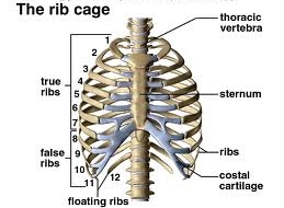
  1. There are 7 sets of true ribs, ribs 1-7
  2. There are 5 sets of false ribs, ribs 8-12
  3. Rib sets 11-12 are considered floating ribs
  4. Ribs move when we breath
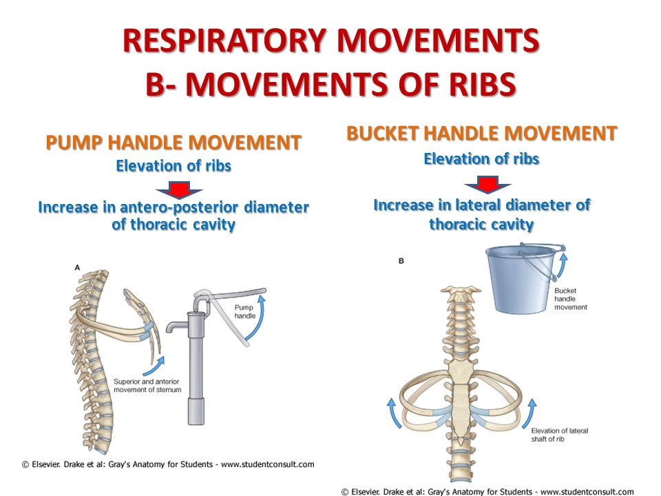
  5. The true ribs are dominated by a pump handle action
  6. The false ribs are dominated by a bucket handle action

Understanding Fact #1

The first 7 sets of ribs directly attach to the sternum anteriorly and to the thoracic spine posteriorly.  This forms a singular unit that is stable and rigid. While this helps to protect the heart and lungs, its' position and motion will dictate the function and orientation of the scapula as well as influence apical lung expansion.

Understanding Fact #2

Rib sets 8-10 are considered false ribs.  These ribs have a posterior attachment to the thoracic spine, but anteriorly they only connect to the sternum through costal cartilage.  This makes them more mobile than the true ribs and allows them to be influenced by our abdominals. Our abdominals help the ribs facilitate the respiratory function of the diaphragm.  This is reflected in the infrasternal angle (ISA). A normal ISA is about 90 degrees, if wider, greater than 100 degrees, it indicates poor abdominal opposition and a diaphragm posturally orientated.  If less than 90 degrees, there is likely an abdominal imbalance driving a similar diaphragm orientation.

Understanding Fact #3

Rib sets 11-12 are also false ribs, but classified as our floating ribs.  These ribs attach posteriorly to the thoracic spine, but do not have an anterior attachment.  These ribs do not serve a significant role in the respiratory process, but are critical for protection of vital organs like the kidneys and the adrenal glands.

Understanding Fact #4-6

rib motions2.jpg

Facts 4-6 are all about normal rib mechanics during respiration.  As we breathe the ribs have to move in such a way to optimize thorax expansion.  All ribs upon inhalation will externally rotate and elevate, anteriorly, and internally rotate and depress, posteriorly.  The opposite occurs during exhalation. However this is only part of the story for optimal thorax expansion. The rest of the story is found at the thoracic spine.

The orientation of the costovertebral joints are different for the upper and lower ribs.  As a result the defining motions within these ribs also differs. The movement of true ribs can be best seen from a lateral view and resembles the motion of a pump handle.  Whereas the false ribs have more of a bucket handle motion and can be seen posteriorly. These normal mechanics both work to best increase the thorax dimensions during inhalation and decrease it with exhalation. This in turn creates a pressurized system that will drive bidirectional airflow.

But how does this influence our movement?

Our movement is dependent upon how we manage this pressure system.  You see, as the arms and legs move, they will change the shape of the thorax and alter the airflow as well as the pressure within it.  As the thorax shape changes so does the orientation of our pelvic innominates and scapulae. So if we have poor rib mechanics or don't manage our thorax pressure well we will begin to compensate and restrict our movement patterns.  Thus it becomes increasingly important to be good pressure managers to avoid these compensatory movement strategies.

The ribs have become vastly under appreciated in our movement health.  They can and do influence multiple body parts as well as systems making them an ideal starting point for almost every injury type.  Hopefully you can appreciate this and now have a better handle on why our ribs matter in the restoration of our movement health.

Stay Well, Stay Strong

Keaton


References:

  1. Hartman, Bill, ALL GAIN, NO PAIN: The Over-40 Man's Comeback Guide to Rebuild Your Body After Pain, Injury, or Physical Therapy.  William Hartman. 2017.
  2. Lee DG. Biomechanics of the thorax - research evidence and clinical expertise. J Man Manip Ther. 2015;23(3):128-38.
  3. Neumann DA. Kinesiology of the Musculoskeletal System, Foundations for Rehabilitation. Mosby; 2010.

Understanding the Airway Part 3: The Diaphragm

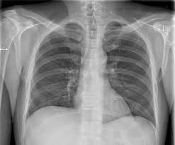
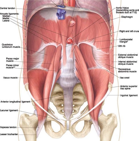
In Parts 1 and 2, we discussed the relationship between the airway and stress on the body and how the upper airway plays into that. In this article, we will discuss the prime mover of the respiratory system, the diaphragm. The diaphragm muscle is especially important, not just because it keeps us alive but also because we use it roughly 25,000 times per day. It then becomes important to know how it functions, and this requires an understanding of anatomy.  Here’s a few important facts about the anatomy of the diaphragm:

thorax.jpeg
  1. The right half of the diaphragm (hemi-diaphragm) is larger, thicker and stronger than the left.

  2. The crura (“legs”) of the diaphragm attach to the bodies and disks of the 1st, 2nd, and 3rd lumbar vertebrae on the right and the 1st and 2nd on the left.

  3. The medial ligaments of the diaphragm cross over the psoas, a muscle which has a hip flexor function.

  4. The position of the right hemi-diaphragm over the liver assists in keeping the right side in its resting domed shape whereas the position of the left hemi-diaphragm under the pericardium assists in keeping the left side in its flattened descended active state.  

  5. The arcuate ligaments of the diaphragm on the lumbar spine can act to “tighten” the back into an arched and loaded position.

  6. During inhalation, the diaphragm descends into a “flattened” shape and during exhalation the diaphragm forms into a domed shape allowing airflow.  

diaphragm and hip flexors.jpg

A few things that we can learn about function of the diaphragm based off of these facts is that the diaphragm is a muscle with a stronger pull on the right.  This means as we breathe 25,000 times per day we may be cranking the spine to the right. The diaphragm is also an important muscle in regards to posture. If the abdominals, especially the internal obliques and transverse abdominus, become weak, the diaphragm may stay descended on both sides into a “flat” and shortened state resulting in potentially tight backs and hip flexors.  When this occurs the diaphragm will assume a more postural stabilizing role rather than its desired respiratory role. We may also notice rib flares, especially on the left due to the position and asymmetry of the diaphragm. This shortened state of the diaphragm makes it weaker as a respiratory muscle because it lacks a normal length tension relationship. Because of this we may end up using accessory muscles of the back and shoulders to inhale which may result in tight shoulders and necks.  Though this doesn’t sound like it should be a problem because you’re just breathing, but you’re doing it 25,000 times per day!

This strongly relates to stress.  The resulting decrease in intra-abdominal pressure results in a “hyper-inflated” state.  This can lower the CO2 in the body which increases the “fight or flight” response resulting in an increased breath rate at rest.  This can also restrict blood flow to the cerebral cortex of the brain, impair gastrointestinal blood flow, promote fatigue and weakness, increase sympathetic adrenal activity, increase anxiety, as well as make you more sensitive to light and sounds.  Like we discussed in Part 2 these are all strongly associated with mouth-breathing.

Now that we’ve seen what inefficient breathing looks like at rest, let’s look at optimal mechanical function of the diaphragm and how a physical therapist can help you out in this regard.  

Optimal mechanical function and power of the diaphragm occurs when the diaphragm is able to go in and out of its resting domed shape and flattened active state on both sides and even be able to alternate in the appropriate conditions.  The diaphragm is mechanically coupled with the abdominals and the rib cage and these all depend on each other for optimal function. This relationship is referred to as the “zone of apposition.” Abdominal disuse can result in flared ribs and a loss of a zone of apposition.  This may appear as “belly breathing.” There’s nothing wrong with belly breathing however if this is the preferred way of respiration, the result can be an elevated stress response and the development of pain syndromes. A physical therapist can help you to restore a zone of apposition by helping you to normalizing resting abdominal tone thereby increasing intra-abdominal pressure and allowing the diaphragm to rest in its domed shape.  In this shape, the diaphragm will function with the abdominal musclature in a piston-like movement allow us to avoid overextending and tightening up the lower back. In turn, this will end up relaxing accessory breathing muscles throughout the body: back, shoulders, and neck. Hence, how you breathe matters, especially since we do it 25,000 times per day!

As always, take care, and breathe easy!

Dave


References

  1. Postural Respiration: An Integrated Approach to Treatment of Patterned Thoraco-Abdominal Pathomechanics. 2000-2016.

  2. Boynton B, Barnas G, Dadmun J, Fredberg J: Mechanical coupling of the rib cage, abdomen, and diaphragm through their area of apposition. J Appl Physiol 70:3,1991.

  3. Cassart M, Pettiaux N, Gevenois PA, Paiva M, Estenne M. Effect of chronic hyperinflation on diaphragm length and surface area. Am J Respir Crit Care Med. 156:504-508, 1997.

  4. Estenne M, Derom E, DeTroyer A. Neck and abdominal muscle activity in patients with severe thoracic scoliosis. Am J Respir Crit Care Med. 1998 Aug;158 (2):452-457.

  5. Goldman M, Mead J: Mechanical interaction between the diaphragm and the rib cage. J Appl Physiol 35:2,1973.9.Hodges P, Gandevia S, Richardson C: Contractions of specific abdominal muscles in postural tasks are affected by respiratory maneuvers. J Appl Physiol 83:3, 1997.10.

  6. Hruska RJ: Influences of dysfunctional respiratory mechanics on orofacial pain. Dent Clin North Am 41:2,1997.

Understanding The NeuroImmune System

neurobrain.jpg

The nervous system is truly the most incredible and adaptable biological system ever created.  The nervous system is an intelligent design composed for bidirectional biological communication to ensure survival.  I was fortunate enough to attend a course, Mobilization of the Neuroimmune System, this weekend to enhance my understanding of how to best recognize and approach a comprised nervous system.

Weekend Takeaways:

  1. The immune system heavily influences the nervous system
  2. Neurodynamic tests assess the physical capacity of the nervous system
  3. Active treatment is more than just moving it also includes breathing and learning.
  4. The type of biological container implicated matters for treatment approach
  5. "Rehab exercises" are not as much about strength as they are about homuncular clarity

In this post I want to explore the first take away to help illustrate briefly how the nervous system and immune system are related.  

The Immune System Heavily Influences The Nervous System:

nervous system.jpg

We got the weekend started by diving deep into the neurobiology of the nervous system and its relationship with the immune system.  Traditionally these two systems have been viewed as separate entities, but that view is now stale, especially when pain enters the picture.  It would now be appropriate to view the nervous system as a neuroimmune organ. Have you ever wondered why you get a whole body ache when you are sick?  Or why your musculoskeletal pain worsens when sick? What about why your pain increases with a weather change? Each of these are due to an elevated neuroimmune system response based on past experiences.  The immune system is a system that knows who you are and will intervene whenever you are not yourself. The immune system is very efficient as there are nearly ten times the amount of immune cells than there are neurons (nerve cells). This is a beautiful and efficient design for protection.

Unfortunately the initial response of the nervous system tends to be inflammation and a painful experience because they are most effective at altering behavior and reducing an imposed threat.  But why does this occur with something as simple as a weather change or going on a walk? Well, the human brain never forgets. It has an amazing capacity to remember and store memories within the hippocampus.  When memories are stored so are the emotions and the environmental context of the time. So when an event or environment is encountered the body will recall memories, the associated emotions, as well as the environmental context to predict an appropriate outcome.  If there was a harmful experience in the past you may trigger an immune response. How? Interestingly enough, your emotions are derived from the amygdala and memories, the hippocampus, both of which are immune system mediators. This means that your past experiences can trigger immune responses and dictate how the nervous system responds.

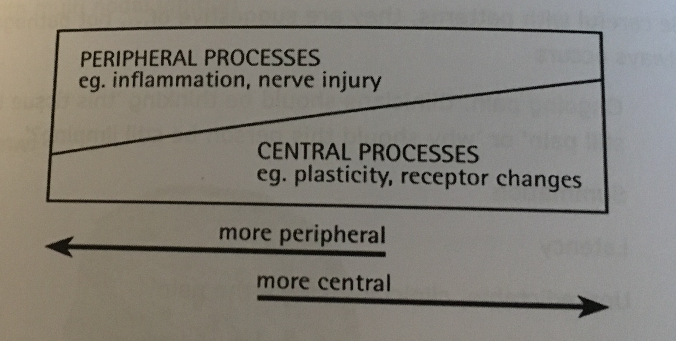
How the nervous system responds is dependent upon the pathobiological mechanism, or how the brain processes the incoming information.  There are two types of mechanisms: pain and tissue. The pain mechanism tends to be related to central processing and viewed as a top-down process.  When this occurs there has become alterations in the central nervous system causing the brain to become too protective with its’ predications. There has become a belief there is more danger than there is in reality.  Danger will evoke F.E.A.R (false evidence appearing real). Remember your emotions can trigger an immune response even if unnecessary. This in turn begins to make your nerves more sensitive and more irritable for the future.  

Tip:

Often you can determine if there is a central processing issue by placing a hot pack on a painful region.  If you think about it from a physiological perspective this should increase inflammation and make you feel WORSE!  So if heat feels good be thankful because the tissue you think is damaged is likely not as bad as you think!

The tissue mechanism is related to peripheral processing, or a bottom-up process.  This is the typical process during an acute injury and follows the predictable stages of healing.  When a tissue is injured inflammation is sent locally to start the healing process. This inflammation triggers the neuron to fire a signal to the brain. If pain is the output, inflammation will be sustained to allow the tissue to heal.  Remember inflammation is a protector. So when inflammation persists it will reduce the firing threshold of the neurons, making it easier for them to be triggered. This prevents you from further tissue damage, but makes you more likely to experience pain with normally non-painful activities.  Interestingly though this is not just an acute scenario. Once you have healed completely, your body will leave some resident immune cells in the previously injured area just in case. This is why your body can remember your pain so well and allow a “relapse” to occur. Remember the brain never forgets and always predicts.

Tip:

We do not have pain receptors!  We have nociceptors that send a signal to the brain where it is decided whether or not pain is the appropriate response.  This is why tissue damage and pain rarely correlate!

FullSizeRender (9).jpg

That being said, deciphering if it is a pain or tissue mechanism is not as simple as placing a hot pack and seeing what happens.  Central and peripheral processes occur simultaneously, but which is the dominant player is the critical aspect. In part 2 of this series I will review how the use of Neurodynamic testing can help to illuminate the physical capacity of the nervous system and guide this clinical reasoning process.

Stay Well, Stay Strong

Keaton

Understanding The Airway Part 2

upper_airway.jpeg

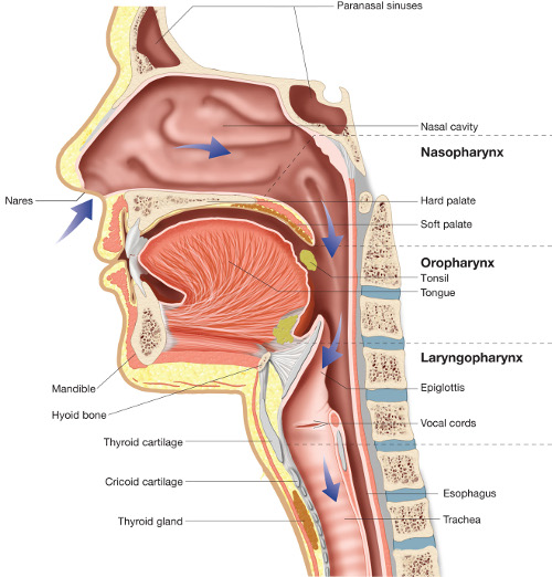
 In Part 1 we discussed the airway and how it relates to the stress response at a general level.  In part 2 we will discuss the airway at the level of the head and neck and how that affects stress and posture.  The primary factors we will look at are Jaw position/bite, tongue position, and the nasal cavities.

The human body is very sensitive to the size of the airway.(1)  The size of the airway dictates the brain’s perception of the environment as being safe or threatening.  A small airway can result in a sensitive stress response as the oxygen demand will more quickly exceed the supply.  Factors such as whether or not a child was breastfed and for how long, when a child was started on hard foods, and if a child uses a pacifier can all be related to the development size of one’s airway.(2,3,4)    First, let’s talk about jaw position.

Jaw Position:

Research has been found that it only takes a two mm shift in jaw position to cause or alleviate sleep apnea.  This is important because sleep apnea, which can lead to a state of low oxygen in the body, is commonly found in individuals with pain complaints.(5,6,7,8)  A state of low oxygen is a massive stressor to the body.  

Tongue Position

If a small shift in jaw position is enough to affect sleep and the airway, tongue size and position is important as well.(9)  Tongues that are restricted because of tongue ties, tend to fall back in the airway easily especially during sleep.(10)  Restricted tongues also affect development of the airway too.  Larger tongues are more likely to fall back and block the airway during sleep.(11,12)  The reason a tongue is large is often linked to how it is being used.  Tongues can become large when allowed to relax and muscle tone is reduced.  However, a tongue that is applying light pressure to the hard palate behind the incisors will actually take up less space and open up the airway.(12)  The caveat is that the tongue cannot do this alone.  It requires nose breathing.

Nasal Cavity

The nasal cavity is extremely important in regards to the airway.  For instance nose breathing in and of itself helps to reduce blood pressure and decrease muscle activity that leads to shoulder and neck tightness.(13,14)  A deviated nasal septum or a hole in the nasal septum is also a potential factor in air way obstruction.

aprilcolumn.png

There are many factors related to airway obstruction and how the airway is related to pain complaints.  In this article we addressed three evidence based factors related to the airway and musculoskeletal pain.  Though it is not always a requirement to have “ideal” tongue posture, jaw position, or nasal cavity, it can be important to consider each of these as possible factors related to pain complaints especially when one has had no results or recurrent episodes of pain complaints.  

As always, take care and breathe easy!

Dave


References

  1. Izuka EN, Feres MF, Pignatari SS. Immediate impact of rapid maxillary expansion on upper airway dimensions and on the quality of life of mouth breathers. Dental Press J Orthod. 2015;20(3):43-9.

  2. Brew BK, Marks GB, Almqvist C, Cistulli PA, Webb K, Marshall NS. Breastfeeding and snoring: a birth cohort study. PLoS ONE. 2014;9(1):e84956.

  3. Page DC. Breastfeeding is early functional jaw orthopedics (an introduction). Funct Orthod. 2001;18(3):24-7.

  4. Howard CR, Howard FM, Lanphear B, et al. Randomized clinical trial of pacifier use and bottle-feeding or cupfeeding and their effect on breastfeeding. Pediatrics. 2003;111(3):511-8.

  5. Nijs J, Loggia ML, Polli A, et al. Sleep disturbances and severe stress as glial activators: key targets for treating central sensitization in chronic pain patients?. Expert Opin Ther Targets. 2017;21(8):817-826.

  6. Köseoğlu Hİ, İnanır A, Kanbay A, et al. Is There a Link Between Obstructive Sleep Apnea Syndrome and Fibromyalgia Syndrome?. Turk Thorac J. 2017;18(2):40-46.

  7. Silva A, Mello MT, Serrão PR, et al. Influence of Obstructive Sleep Apnea in the Functional Aspects of Patients With Osteoarthritis. J Clin Sleep Med. 2018;14(2):265-270.

  8. Martinot JB, Borel JC, Cuthbert V, et al. Mandibular position and movements: Suitability for diagnosis of sleep apnoea. Respirology. 2017;22(3):567-574.

  9. Yoon AJ, Zaghi S, Ha S, Law CS, Guilleminault C, Liu SY. Ankyloglossia as a risk factor for maxillary hypoplasia and soft palate elongation: A functional - morphological study. Orthod Craniofac Res. 2017;20(4):237-244.

  10. Harvey R, O'brien L, Aronovich S, et al. Friedman tongue position and cone beam computed tomography in patients with obstructive sleep apnea. Laryngoscope Investig Otolaryngol. 2017;2(5):320-324.

  11. Barrera JE, Pau CY, Forest VI, Holbrook AB, Popelka GR. Anatomic measures of upper airway structures in obstructive sleep apnea. World J Otorhinolaryngol Head Neck Surg. 2017;3(2):85-91.

  12. Hwang DM, Lee JY, Choi YJ, Hwang CJ. Evaluations of the tongue and hyoid bone positions and pharyngeal airway dimensions after maxillary protraction treatment. Cranio. 2018;:1-9.

  13. Gelardi M, Abbattista G, Quaranta VN, et al. Standardization procedure for the nasal nitric oxide measurement method using Niox MINO® and the tidal-breathing technique with velum-closure. J Biol Regul Homeost Agents. 2016;30(3):853-858.

  14. Ip MS, Lam B, Chan LY, et al. Circulating nitric oxide is suppressed in obstructive sleep apnea and is reversed by nasal continuous positive airway pressure. Am J Respir Crit Care Med. 2000;162(6):2166-71.

Understanding The Airway

stress and breath.png

In the physiological hierarchy of needs, the body’s number one requirement is air.(1)  Without air the body would die within minutes so it makes sense that the body will utilize as many strategies as necessary to maintain a functional oxygen level in the body.  When the brain perceives that it is under stress, the demand for oxygen increases and the fight or flight response is triggered. This involves increased heart rate, increased blood flow to working muscle, and decreased blood flow to systems that are not required for short term protection such as digestion and the immune system.(2)  This stress response is also reflected in posture and muscle activation.  A very clear example of this is the startle response which involves the body attempting to escape a stressor.(3)  Another example is watching individuals at the end of a marathon.  They inevitably begin to arch their back and push out their chest as they try to pull in more air to keep going.  These are all normal reflexes that the body exhibits to take care of itself.

The stress response is efficient during short term stressors.  Unfortunately, in today’s society we are constantly bombarded with mild stressors which involve everything from going to work, paying bills, and managing home and technology.(4,5)  This puts an individual's body in a state of constant mild distress yielding a prolonged stress response.6  The brain adapts to this stress response and creates a "new" normal "resting" state, one that is resemblant of the marathon runner.  The lower abdominal muscles become less active allowing the low back to tighten and arch. This is an example of a postural change striving to get more air in the body.  Paradoxically, this posture is one of hyperinflation, or too much air in the lungs. This increases blood pH levels, which in turn increases the perceived threat. This is the hallmark of an inefficient breather.  Oxygen utilization is low when stressed. To get more "useful" air in the old air needs to come out more efficiently during exhalation. Over time, if “stuck” in this or an associated survival posture it can lead to musculoskeletal pain complaints such as low back, neck, or even shoulder pain.  The goal becomes to build a body with the capacity to assume multiple postures without getting stuck. The body should be able to go in and out of postures if it is no longer under threat. The most efficient way to reduce a threat is with a respiration emphasis. Respiratory patterns train the brain to decrease the state of threat on the body.  

This concludes Part 1 of the airway.  Though it is not necessarily a requirement to have “perfect” posture or stress management, breathing is something we do thousands of times a day and can be the first step to improving both of these.  

As always, take care and breathe easy!

Dave


References

  1. Maslow, A. H. (1943). A theory of human motivation. Psychological Review, 50(4), 370-396.

  2. Cannon, Walter (1932). Wisdom of the Body. United States: W.W. Norton & Company. ISBN 0393002055.

  3. Solomon; Schmidt (1990). "13". In Carol, Field. Human Anatomy & physiology (2 ed.). Saunders College Publishing. p. 470. ISBN 0-03-011914-6.

  4. Schneider S, Schmitt H, Zoller S, Schiltenwolf M. Workplace stress, lifestyle and social factors as correlates of back pain: a representative study of the German working population. Int Arch Occup Environ Health. 2005;78(4):253-69.

  5. Konietzny K, Chehadi O, Streitlein-böhme I, Rusche H, Willburger R, Hasenbring MI. Correction to: Mild Depression in Low Back Pain: the Interaction of Thought Suppression and Stress Plays a Role, Especially in Female Patients. Int J Behav Med. 2017.

  6. Robert M. Sapolsky. Why Zebras Don't Get Ulcers: An Updated Guide To Stress, Stress Related Diseases, and Coping. 2nd Rev Ed, April 15, 1998. W. H. Freeman ISBN 978-0-7167-3210-5

Self Myofascial Release: Are You Being Effective AND Efficient?

SMR1.jpg

One of the hot topics within the physical therapy and performance sectors is Self Myofascial Release (SMR).   Today, it seems the debate revolves around weather or not a tissue is really being influenced or if it is wasted time and energy.  This is not a road I care to travel down because I am unlikely to actually change a viewpoint with this writing. However, if you do choose to perform SMR I want you to be as successful as possible.  I want to share five ways both effectiveness and efficiency can be improved.

Reduce surface area

The traditional foam roller is very broad and contacts a larger surface area.  This is not a bad thing, but with increased surface area there is reduced peak pressure applied.  However if you find an exquisitely tender point within the fascial system you may be better served to replace the foam roller with a smaller implement such as a mobility sphere, lacrosse ball, softball, etc.  By doing so, peak pressure is increased and acute tissue adaptation is more likely. However it is unknown how much pressure is created during an SMR technique while using a foam roller. The current biological research shows that for a soft tissue to deform, a moderate to large amount of pressure is needed.  Increased levels of pressure is believed to stimulate mechanoreceptors within the fascial system to yield the biological response desired in the targeted tissue. Thus, to achieve an optimal response, it is logical to consider reducing the surface area.

Move in 360 degrees

Moving away from the foam roller and switching to a sphere of some sort also allows you to manipulate the tissue in multiple planes, or rather cover all 360 degrees.  Tissue adhesions/trigger points don’t occur uniformly. For simplicity, think of a healthy tissue as uncooked spaghetti. Just like uncooked spaghetti, muscle fibers tend to run parallel to one another and intentionally influence the position of multiple fibers.  This is contrasted with an unhealthy tissue full of adhesions, which is more like cooked spaghetti. You try and pull a single noodle out and it will increase the stress applied to a large quantity of the noodles. It seems nearly impossible to free a single noodle if you only try to pull in one direction.  Now apply this to an SMR technique, what happens when only one plane of motion is addressed? Limited success.  The fascial system cannot be freed by addressing only one direction. Rolling in all directions will give a better chance of making a worthwhile adaptation.

Add Active motion

Another useful angle is very similar to the pin and stretch technique.  Some of the most effective forms of manual therapy occur when a muscle is shortened, contact is placed on a palpable adhesion, and then the muscle is lengthened through its’ range of motion.  Similarly if a restriction is found in a muscle regardless of the implement, foam roller or sphere, if the tissue is pinned at an adhesion and then that SAME MUSCLE is moved back and forth through its range of motion you can achieve a similar result.

Move Adjacent musculature

Moving the adjacent musculature is a fourth modification that can be made.  It too builds off of the pin and stretch concept. Myofascial research shows that our muscles are meant to slide and glide past one another.  This is a sign of efficient movement. However some adhesion formations will restrict this muscle play and impact movement patterns. Thus if a muscle is again pinned at an adhesion and the ADJACENT MUSCLE is moved through its full motion, muscle play can be restored to an optimal state.  

Create Compound Context

The final technique to improve efficiency and effectiveness hangs its hat on the fact that the effects of foam rolling are very short lasting.  This is why people feel like they need to continuously “roll out.” If an SMR technique is paired with an exercise emphasizing the mobility of the same structure the effect will be greater.  The rationale or belief is that SMR increases the available mobility, which increases the neurological window for retraining a movement pattern. Thus, performing back to back exercises or incorporating SMR into a circuit can create neurological priming to a tissue and thus allow for a greater adaptation when the tissue is subsequently loaded.  This forces muscular adaptation. The result, more resilience and sustainability within a tissue.

Try any or all of these five techniques the next time you perform a Self Myofascial Release technique to improve both your effectiveness and efficiency.  This will allow you to redirect more of your time and energy into your exercises to create longer lasting adaptations that will keep you healthy!

Stay Well, Stay Strong,

Keaton

The Sportsman: A Journey Together

Timing_Success.jpg

Sports are the most captivating scenes in America.  We can all identify the iconic sportsmen of our childhood and we all know what it means when we hear, “I wanna be like-Mike”.  However, most of us fall short and recognize these sportsman as the lucky few who were blessed with God-given talent that reached super-stardom, effortlessly.  This is not true though, the journey of a sportsman is anything but effortless.  No journey is effortless.  And no journey occurs in isolation.    

In reflection of the sportsman journey, I see the journey of a patient.  The sportsman is usually part of a team who cohesively works together to achieve a common goal:  winning.  A team struggles as well as finds success as a singular unit.  Similarly a patient is part of the rehabilitation team striving to succeed at restoring functional capacity.  However there are often roadblocks that slow this process.  Identification of how the sports team and rehab team constructs are similar can allow us to begin to see where our roadblocks may be developing and how we can overcome them.

So here’s the breakdown:  coaches = physical therapists, players = patients, opposition = movement/behavioral dysfunction, and the officials = our pain.  Recognizing that not only are we a part of a team, but we carry a specific role is powerful.

Let’s dive into each of these to learn how to overcome our roadblocks.  

dickbarvetta.jpg

Distinguishment between the opposition and the official is essential.  Far too often we hang our hat on “It’s my pain’s fault.”  Why?  It’s always easier to blame someone or something else.  Just like in basketball, it is easier to say “the refs cost us the game.”  However we know this is not the case.  The officials are present to ensure we play be the rules and to alert us if we do anything wrong just like our pain does.  To not get the whistle blown on us again we have to change our behavior.  However, this can be incredibly difficult, but it’s not just you, even elite athletes have ordinary habits they struggle to overcome.  A recent sport psychology article identified three primary drivers of behavioral change in elite athletes:  credibility, reliability, and intimacy.  However the interesting part is that these three primary drivers are not about the elite athlete, but rather their coach.     

This is no different in physical therapy.  A patient and physical therapist must not only trust one another, but they have to trust the process.  This starts with effective communication from the physical therapist.  An explanation of a patient’s condition or procedure allows the credibility of the physical therapist to be highlighted.  Providing knowledge and resources alike ensures a person they are in good hands.  The outcome is often improved compliance in their behavioral modifications.  Knowledge can be the liberator needed by a patient to break them free of a movement dysfunction.

However sometimes more than one explanation is needed to help break a chronic movement/behavioral pattern.  Being a reliable and consistent resource is also critical.  Often a person leaves a session feeling good about their exercise program as well as her behavior changes, but return ill-confident in their performance.  Utilizing repeated exposures and reassuring a person can go along way.  And just because a person struggles doesn’t mean they are doing something wrong and need “more change”.  It is quite the contrary, at the point of struggle is the start of learning, so consistency in their performance becomes essential.  The physical therapist should highlight this notion and coach each person through their exercises as they learn how to regain control of their movement patterns.  

That being said though sometimes frustration sets in and hinders performance.  Early recognition of this by the physical therapist is important.  This reflects the intimacy of the relationship between a patient and physical therapist.  Being able to pick up on differences in mood, verbal language, and/or body language can mitigate the frustration roadblock and allow for a consistent restoration of their movement and resolution of symptoms.  

In the end, a physical therapist is in the coaches seat.  Their influence drives and guides the success of the players.  The role of the coach is to maintain focus on overcoming the opposition and not the officials.  To do so hangs on the three primary drivers of behavioral change:  credibility, reliability, and intimacy.  

Stay Well,

Keaton


References:  

  1. Butler DS, Moseley GL, Sunyata. Explain Pain 2nd Edn. Noigroup Publications; 2013.

  2. Halson SL, Lastella M. Amazing Athletes With Ordinary Habits: Why Is Changing Behavior So Difficult?. Int J Sports Physiol Perform. 2017;12(10):1273-1274.

What’s Driving Your Foot Pain?

plantarfascia.jpg

Heel pain or foot pain can be one of the most debilitating conditions for an individual.  We are designed to operate in a bipedal state. Whether we are standing, walking, running, or jumping, we are meant to be on our feet. But what happens when we experience heel or foot pain and we can’t tolerate these activities?  

Often blamed is the plantar fascia, the broad triangular connective tissue, anchored at the medial aspect of the calcaneus (heel) that spans down to the toes.  Its role is to act as our first shock absorber that dissipates ground reaction forces as we move throughout our environment.  It secures the infrastructure of the foot by supporting the medial longitudinal arch, making it an essential feature for optimal foot health.  But I am here to tell you, plantar fasciitis, is not just a foot issue.  Actually, it rarely is.  Only about five percent of cases don’t resolve conservatively and require surgical intervention.

The plantar fascia plays a big role in our gait efficiency, but so does our pelvis and thorax.  Do these structures function independently of each other?  No, but they do influence one another.  Our proximal dysfunction can actually drive our foot symptoms.  We tend to hurt at our weakest point and not at the point of dysfunction.  This is where most treatments are lost.  It hurts at “X” so let me rub and massage “X.”  Treatment becomes myopically focused on tissue/structure and “damage” rather than addressing the underlying mechanism.  

We need to understand pain isn’t bad and it doesn’t necessarily equate to the degree of tissue damage.  Pain is a perceptual response to a threat.  It protects our bodies by telling us something isn’t right and we need to change a behavior to reduce said threat. 

Now this doesn’t mean we ignore the foot or negate manual therapy application.  It means there has to be an appreciation for the interconnected nature of our body to guide intervention.  

This is why it is important we take a step back from the typical local perspective and appreciate the global perspective that yields a lot more as we recognize both intrinsic and extrinsic risk factors for plantar fasciitis.

It starts at a societal level. Today is all about being able to do more, when in reality we don’t have the capacity to do so.  We, as a people don’t know how to handle more because we don’t have effective stress management strategies. The literature is reporting the highest levels of chronic stress, fatigue, and anxiety and with this comes more chronic flight/fight, or sympathetic nervous system activity.  This is the same part of the nervous system that becomes active when we experience pain.  Coincidence? I don't think so.  Our bodies can't distinguish between stressors. They simply interpret and react to ensure survival.  First order of importance when stressed, shift metabolic resources and assume a survival posture.  It is this stress response though that influences the intrinsic risk factors associated with plantar fasciitis:  big toe mobility, ankle dorsiflexion, calf muscle “tightness”, and hip motor control.  

LEmechanics.jpg

As our posture changes to a more extended form, we will see backs that are over extended, rib cages elevated, pelvic girdles dropped forward, and calves with an increase in tone.  All of this will inherently limit our movement variability and subsequently overload our feet each step we take.  This survival posture, thus increases the metabolic demand of the plantar fascia.  It alters the function of the big toe and ultimately the windlass mechanism, which is crucial in preventing our arch from collapsing during the mid-stance of gait.  As we achieve a mid-stance position, our plantar fascia tightens thereby pulling the big toe into extension and enhancing ankle dorsiflexion.  When efficient, it will prevent excessive foot pronation (arch collapse), which is present in 81-86% of plantar fasciitis cases.  It truly is a chain effect.   

The chain continues as we move up to the hip.  Our motor control of the mid-stance position is a reflection of foot and hip integration.  However control becomes increasingly difficult the longer we remain overly extended.  This posture poorly positions hip musculature and unfortunately sets the stage for compensation at the first opportunity, the foot.  Our foot is not only the first intimate interaction with the environment, it is our first chance to respond to it.  However, if we don’t have the proximal orientation of the hip and pelvis, we can’t expect our foot to respond efficiently.  Don’t believe the hip has an influence?  Try this. Stand-up without shoes and watch what happens when we squeeze/tighten our butt.  THE ARCHES LIFT!  Every bad foot can be controlled by a good hip.  Our hips and pelvis have a powerful influence over our feet and must not be neglected in the rehabilitation process.

So here is the relevance of this matter.  

Currently 10% of the general population will develop plantar fasciitis in their lifetime and it will most often occur during occupational years.  Given that these years have proven to be the most stressful, we will be more prone to living our lives in a survival posture and thereby overloading our feet.  If left unchecked, we could miss work, lose out on wages, or experience a reduction in work productivity.  Outside of our work field, it could generate fear-avoidance behaviors to avoid the pain experience.  In turn, we reinforce bad behavioral choices and potentially elevate our stress.  Now we are in a pain-cycle searching for a way out.  

The way out of this cycle is not only addressing the aforementioned intrinsic risk factors through position, but also by addressing our extrinsic risk factors.  

As mentioned earlier, pain is an indication we need to have a behavioral change, most notably our extrinsic factors:  poor footwear, prolonged occupational weight bearing, and large increases in activity level.

Heels.jpeg

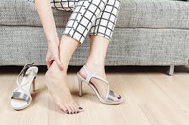
All extrinsic factors are easily modifiable, but usually hard to adhere too, especially when it comes to our footwear.  Poor unsupported footwear such as; heels, sandals, flats, and some work boots don’t provide enough sensory input and end up reinforcing poor intrinsic factor development.  Despite knowing the benefits of footwear, people still struggle for various reasons:  work, financial, etc.  This is why orthotics are often sought after by the public.  Belief is that an orthotic can be the fix for poor footwear.  This may or may not be the case, but regardless it shouldn’t be the first line.  Establishment of an effective home exercise program has been shown to be just as effective when addressing the underlying mechanisms.  This should be our first line.  

The physical stress of being on our feet also cannot be overlooked, which is why prolonged occupational weight bearing is a risk factor that needs to be considered.  If we are on our feet 40+ hours week for 20 or 30 years straight, we are accumulating a lot foot stress.  Research shows walking just one mile a foot can endure nearly 60 tons of stress.  We need to have efficient foot posture and mechanics to withstand this amount of stress over the long haul.

Relating to tissue overload, large increases in our activity level can also increase our susceptibility to plantar fasciitis.  Although our bodies are adaptable they are more concerned with survival.  So if we make too large of a jump in our exercise regimen, intensity or duration, our body will let us know.  Thus, a strategic application of physical activity to control the purposeful stress of exercise is needed to allow the body to best adapt without any repercussions.  

So to recap, plantar fasciitis is not just a foot issue.  It is a multifactorial issue that demands respect of the body’s natural design.  Proximal dysfunction is most likely the offender and the foot the victim.  The success of treatment hangs on the ability to identify and address all intrinsic and extrinsic factors that are working against our feet.  And the good news?  Most of these risk factors are modifiable.  We are in control.     

Stay Well,
Keaton

References:

  1. Ratey JJ. Spark, The Revolutionary New Science of Exercise and the Brain. Little, Brown; 2013.

  2. Rathleff MS, Mølgaard CM, Fredberg U, et al. High-load strength training improves outcome in patients with plantar fasciitis: A randomized controlled trial with 12-month follow-up. Scand J Med Sci Sports. 2015;25(3):e292-300.

  3. Sapolsky RM. Why Zebras Don't Get Ulcers, An Updated Guide to Stress, Stress-related Diseases, and Coping. 2004.

  4. Tahririan MA, Motififard M, Tahmasebi MN, Siavashi B. Plantar fasciitis. J Res Med Sci. 2012;17(8):799-804.

  5. Thompson JV, Saini SS, Reb CW, Daniel JN. Diagnosis and management of plantar fasciitis. J Am Osteopath Assoc. 2014;114(12):900-6.

Failure and the Weight Loss Resolution

Why are you likely to fail? You should start by asking yourself what it is that you want to achieve, and what are you willing to do to reach your goal. Once you have decided what your end goal is, the next step is developing a detailed plan because a goal without a plan is just a wish. This may include a specific diet, exercise plan, lifestyle changes, etc. Once you have made a road map for yourself, you are more likely to follow through with what you set out to accomplish.

It is also important to bring your friends and family into your weight loss journey. Some fall short of their goals because they made a promise solely to themselves. When left to our own resources, we have no problem letting ourselves down. If you promise someone that you will be there for them, you are far more likely to follow through. The same idea applies to weight loss. If you profess your path to weight loss to someone else, along with specific goals and time frames, you are more likely to succeed.

The keys to a good weight loss strategy:

  • Setting achievable, realistic goals
  • Developing a specific plan
  • Being persistent
  • Holding yourself accountable for setbacks
  • Involving others

Spring time has finally won!

We are over the long, dreary, cold days of winter.  Lawn mowers spring to life, wheel barrows are full of mulch, and everywhere you look, people are shoveling, stooping, and planting.  For most of us, our backs and knees are not as forgiving of an idle winter season as our flower beds.

Following an afternoon’s pleasure in the yard, we are reminded that we overindulged in an activity of which we were unaccustomed, and now we are suffering in our backs, knees, and all over our body.  

In a recent Gallop pole of 2,000 adults, nearly half suffer from back pain and half of those do so as a result of gardening.  Is that really surprising though?  We have spent the last several months inside, away from these activities, and perhaps away from any activity.  We are out of “shape,” and our muscles have become tighter and weaker.  We haven’t done anything to address those nagging aches and pains from last year.  In the spring rush of enthusiasm we then stay at tasks for hours bent over.  We lift heavy loads.  We twist, kneel, stand, and overreach.

So what’s a gardener to do?  The first and most important step is to give your body the same consideration you give your garden by spending a little time planning and preparing.

  1. Prepare:  Those nagging or recurring back or knee problems that limited you last year will still be with you if you haven’t done anything about them.   Just as athletes rehab their injuries and train in the off season, gardeners should do the same.  A comprehensive examination by a skilled physician is the first step to identifying the root cause of your pain and dysfunction.  The prescription will involve starting an appropriate exercise program of focused stretching and strengthening, or perhaps an injection of your arthritic knee may be needed.  Additionally, a  well designed, “preseason” training program of stretching and strengthening will often prevent or limit soreness or new problems. 

  2. Adjust: If you have back and or knee problems, raise the height of your flower beds or sit on a stool.

  3. Warm-up:  Begin every gardening session with some stretches provided by your therapist.

  4. Tools: Use the right tools, whenever possible use long handled, lightweight tools.
      
  5. Work Smart:  Avoid working in a single position more than 20 minutes.  For example, weed for a while, then prune for a while, now plant a few bulbs, etc…

  6. Bend Your Knees: To lift with ease you need to bend your knees. Plan your lifts.  If it looks too heavy ask for help.  If you do lift keep you back straight and lift with you legs.  Always carry the load close to your body.

  7. Water: Drink Lots of Water. Working muscles build up toxins.  Water helps flush them out and limits post activity soreness.

Back to school

A backpack is a great way of carrying the books, binders, supplies, lunches, gym clothes, and everything else needed for school. But, in order to carry all of those things backpacks have become larger and larger... but have they become too large and too heavy? How much is safe for your child to carry?


The American Physical Therapy Association recommends that your child's backpack should never exceed 10-15% of their body weight, although most packs are often close to 30-45% of their weight. When stresses placed on the spine exceed its ability to absorb them, spinal imbalances - and injury- will happen.  A heavy backpack, for example, can pull a child backward, causing them to compensate by either bending forward or arching their back. Slinging a backpack over one shoulder can also cause a child to lean too far to the other side to offset the weight. Over time, this overcompensation can lead to poor posture, muscle strain and pain in the back, neck and shoulders. Backpacks with tight, narrow straps that dig into the skin can cause tingling, numbness, and weakness in the arms and hands.

What can you do?

  • Pack lightly.
  • Organize - heavier items closest to the back and use the compartments.
  • Use both shoulder straps, make sure they are wide and padded.
  • Tighten the straps so the backpack sits close to the back and in the middle.
  • Use a locker, don't care everything at one time.
  • Squat down, bend at the knees not the waist.
  • Encourage your child to tell you if they are experiencing pain.
  • Schedule an appointment with a doctor if they do experience any pain.