respiration

Getting A Handle On Our Ribs

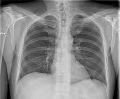
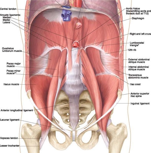
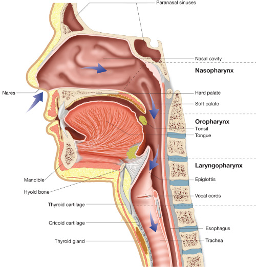
We have recently wrote about the respiratory system: from the upper respiratory airway down to the primary respiratory muscle, the diaphragm.  But what about the protective rib cage? Our ribs play a critical role in not only respiration, but in movement, too.

Let's get a better handle on our ribs.
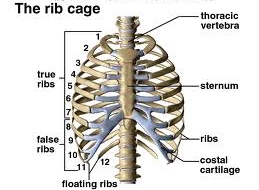
ribcage.jpg

Rib facts:

  1. There are 7 sets of true ribs, ribs 1-7
  2. There are 5 sets of false ribs, ribs 8-12
  3. Rib sets 11-12 are considered floating ribs
  4. Ribs move when we breath
  5. The true ribs are dominated by a pump handle action
  6. The false ribs are dominated by a bucket handle action

Understanding Fact #1

The first 7 sets of ribs directly attach to the sternum anteriorly and to the thoracic spine posteriorly.  This forms a singular unit that is stable and rigid. While this helps to protect the heart and lungs, its' position and motion will dictate the function and orientation of the scapula as well as influence apical lung expansion.

Understanding Fact #2

Rib sets 8-10 are considered false ribs.  These ribs have a posterior attachment to the thoracic spine, but anteriorly they only connect to the sternum through costal cartilage.  This makes them more mobile than the true ribs and allows them to be influenced by our abdominals. Our abdominals help the ribs facilitate the respiratory function of the diaphragm.  This is reflected in the infrasternal angle (ISA). A normal ISA is about 90 degrees, if wider, greater than 100 degrees, it indicates poor abdominal opposition and a diaphragm posturally orientated.  If less than 90 degrees, there is likely an abdominal imbalance driving a similar diaphragm orientation.

Understanding Fact #3

Rib sets 11-12 are also false ribs, but classified as our floating ribs.  These ribs attach posteriorly to the thoracic spine, but do not have an anterior attachment.  These ribs do not serve a significant role in the respiratory process, but are critical for protection of vital organs like the kidneys and the adrenal glands.

Understanding Fact #4-6

rib motions2.jpg

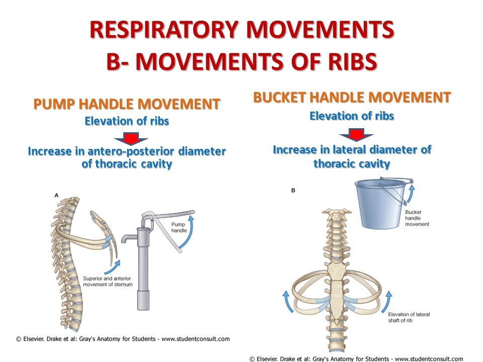
Facts 4-6 are all about normal rib mechanics during respiration.  As we breathe the ribs have to move in such a way to optimize thorax expansion.  All ribs upon inhalation will externally rotate and elevate, anteriorly, and internally rotate and depress, posteriorly.  The opposite occurs during exhalation. However this is only part of the story for optimal thorax expansion. The rest of the story is found at the thoracic spine.

The orientation of the costovertebral joints are different for the upper and lower ribs.  As a result the defining motions within these ribs also differs. The movement of true ribs can be best seen from a lateral view and resembles the motion of a pump handle.  Whereas the false ribs have more of a bucket handle motion and can be seen posteriorly. These normal mechanics both work to best increase the thorax dimensions during inhalation and decrease it with exhalation. This in turn creates a pressurized system that will drive bidirectional airflow.

But how does this influence our movement?

Our movement is dependent upon how we manage this pressure system.  You see, as the arms and legs move, they will change the shape of the thorax and alter the airflow as well as the pressure within it.  As the thorax shape changes so does the orientation of our pelvic innominates and scapulae. So if we have poor rib mechanics or don't manage our thorax pressure well we will begin to compensate and restrict our movement patterns.  Thus it becomes increasingly important to be good pressure managers to avoid these compensatory movement strategies.

The ribs have become vastly under appreciated in our movement health.  They can and do influence multiple body parts as well as systems making them an ideal starting point for almost every injury type.  Hopefully you can appreciate this and now have a better handle on why our ribs matter in the restoration of our movement health.

Stay Well, Stay Strong

Keaton


References:

  1. Hartman, Bill, ALL GAIN, NO PAIN: The Over-40 Man's Comeback Guide to Rebuild Your Body After Pain, Injury, or Physical Therapy.  William Hartman. 2017.
  2. Lee DG. Biomechanics of the thorax - research evidence and clinical expertise. J Man Manip Ther. 2015;23(3):128-38.
  3. Neumann DA. Kinesiology of the Musculoskeletal System, Foundations for Rehabilitation. Mosby; 2010.

Understanding the Airway Part 3: The Diaphragm

In Parts 1 and 2, we discussed the relationship between the airway and stress on the body and how the upper airway plays into that. In this article, we will discuss the prime mover of the respiratory system, the diaphragm. The diaphragm muscle is especially important, not just because it keeps us alive but also because we use it roughly 25,000 times per day. It then becomes important to know how it functions, and this requires an understanding of anatomy.  Here’s a few important facts about the anatomy of the diaphragm:

thorax.jpeg
  1. The right half of the diaphragm (hemi-diaphragm) is larger, thicker and stronger than the left.

  2. The crura (“legs”) of the diaphragm attach to the bodies and disks of the 1st, 2nd, and 3rd lumbar vertebrae on the right and the 1st and 2nd on the left.

  3. The medial ligaments of the diaphragm cross over the psoas, a muscle which has a hip flexor function.

  4. The position of the right hemi-diaphragm over the liver assists in keeping the right side in its resting domed shape whereas the position of the left hemi-diaphragm under the pericardium assists in keeping the left side in its flattened descended active state.  

  5. The arcuate ligaments of the diaphragm on the lumbar spine can act to “tighten” the back into an arched and loaded position.

  6. During inhalation, the diaphragm descends into a “flattened” shape and during exhalation the diaphragm forms into a domed shape allowing airflow.  

diaphragm and hip flexors.jpg

A few things that we can learn about function of the diaphragm based off of these facts is that the diaphragm is a muscle with a stronger pull on the right.  This means as we breathe 25,000 times per day we may be cranking the spine to the right. The diaphragm is also an important muscle in regards to posture. If the abdominals, especially the internal obliques and transverse abdominus, become weak, the diaphragm may stay descended on both sides into a “flat” and shortened state resulting in potentially tight backs and hip flexors.  When this occurs the diaphragm will assume a more postural stabilizing role rather than its desired respiratory role. We may also notice rib flares, especially on the left due to the position and asymmetry of the diaphragm. This shortened state of the diaphragm makes it weaker as a respiratory muscle because it lacks a normal length tension relationship. Because of this we may end up using accessory muscles of the back and shoulders to inhale which may result in tight shoulders and necks.  Though this doesn’t sound like it should be a problem because you’re just breathing, but you’re doing it 25,000 times per day!

This strongly relates to stress.  The resulting decrease in intra-abdominal pressure results in a “hyper-inflated” state.  This can lower the CO2 in the body which increases the “fight or flight” response resulting in an increased breath rate at rest.  This can also restrict blood flow to the cerebral cortex of the brain, impair gastrointestinal blood flow, promote fatigue and weakness, increase sympathetic adrenal activity, increase anxiety, as well as make you more sensitive to light and sounds.  Like we discussed in Part 2 these are all strongly associated with mouth-breathing.

Now that we’ve seen what inefficient breathing looks like at rest, let’s look at optimal mechanical function of the diaphragm and how a physical therapist can help you out in this regard.  

Optimal mechanical function and power of the diaphragm occurs when the diaphragm is able to go in and out of its resting domed shape and flattened active state on both sides and even be able to alternate in the appropriate conditions.  The diaphragm is mechanically coupled with the abdominals and the rib cage and these all depend on each other for optimal function. This relationship is referred to as the “zone of apposition.” Abdominal disuse can result in flared ribs and a loss of a zone of apposition.  This may appear as “belly breathing.” There’s nothing wrong with belly breathing however if this is the preferred way of respiration, the result can be an elevated stress response and the development of pain syndromes. A physical therapist can help you to restore a zone of apposition by helping you to normalizing resting abdominal tone thereby increasing intra-abdominal pressure and allowing the diaphragm to rest in its domed shape.  In this shape, the diaphragm will function with the abdominal musclature in a piston-like movement allow us to avoid overextending and tightening up the lower back. In turn, this will end up relaxing accessory breathing muscles throughout the body: back, shoulders, and neck. Hence, how you breathe matters, especially since we do it 25,000 times per day!

As always, take care, and breathe easy!

Dave


References

  1. Postural Respiration: An Integrated Approach to Treatment of Patterned Thoraco-Abdominal Pathomechanics. 2000-2016.

  2. Boynton B, Barnas G, Dadmun J, Fredberg J: Mechanical coupling of the rib cage, abdomen, and diaphragm through their area of apposition. J Appl Physiol 70:3,1991.

  3. Cassart M, Pettiaux N, Gevenois PA, Paiva M, Estenne M. Effect of chronic hyperinflation on diaphragm length and surface area. Am J Respir Crit Care Med. 156:504-508, 1997.

  4. Estenne M, Derom E, DeTroyer A. Neck and abdominal muscle activity in patients with severe thoracic scoliosis. Am J Respir Crit Care Med. 1998 Aug;158 (2):452-457.

  5. Goldman M, Mead J: Mechanical interaction between the diaphragm and the rib cage. J Appl Physiol 35:2,1973.9.Hodges P, Gandevia S, Richardson C: Contractions of specific abdominal muscles in postural tasks are affected by respiratory maneuvers. J Appl Physiol 83:3, 1997.10.

  6. Hruska RJ: Influences of dysfunctional respiratory mechanics on orofacial pain. Dent Clin North Am 41:2,1997.

Understanding The Airway Part 2

upper_airway.jpeg

 In Part 1 we discussed the airway and how it relates to the stress response at a general level.  In part 2 we will discuss the airway at the level of the head and neck and how that affects stress and posture.  The primary factors we will look at are Jaw position/bite, tongue position, and the nasal cavities.

The human body is very sensitive to the size of the airway.(1)  The size of the airway dictates the brain’s perception of the environment as being safe or threatening.  A small airway can result in a sensitive stress response as the oxygen demand will more quickly exceed the supply.  Factors such as whether or not a child was breastfed and for how long, when a child was started on hard foods, and if a child uses a pacifier can all be related to the development size of one’s airway.(2,3,4)    First, let’s talk about jaw position.

Jaw Position:

Research has been found that it only takes a two mm shift in jaw position to cause or alleviate sleep apnea.  This is important because sleep apnea, which can lead to a state of low oxygen in the body, is commonly found in individuals with pain complaints.(5,6,7,8)  A state of low oxygen is a massive stressor to the body.  

Tongue Position

If a small shift in jaw position is enough to affect sleep and the airway, tongue size and position is important as well.(9)  Tongues that are restricted because of tongue ties, tend to fall back in the airway easily especially during sleep.(10)  Restricted tongues also affect development of the airway too.  Larger tongues are more likely to fall back and block the airway during sleep.(11,12)  The reason a tongue is large is often linked to how it is being used.  Tongues can become large when allowed to relax and muscle tone is reduced.  However, a tongue that is applying light pressure to the hard palate behind the incisors will actually take up less space and open up the airway.(12)  The caveat is that the tongue cannot do this alone.  It requires nose breathing.

Nasal Cavity

The nasal cavity is extremely important in regards to the airway.  For instance nose breathing in and of itself helps to reduce blood pressure and decrease muscle activity that leads to shoulder and neck tightness.(13,14)  A deviated nasal septum or a hole in the nasal septum is also a potential factor in air way obstruction.

aprilcolumn.png

There are many factors related to airway obstruction and how the airway is related to pain complaints.  In this article we addressed three evidence based factors related to the airway and musculoskeletal pain.  Though it is not always a requirement to have “ideal” tongue posture, jaw position, or nasal cavity, it can be important to consider each of these as possible factors related to pain complaints especially when one has had no results or recurrent episodes of pain complaints.  

As always, take care and breathe easy!

Dave


References

  1. Izuka EN, Feres MF, Pignatari SS. Immediate impact of rapid maxillary expansion on upper airway dimensions and on the quality of life of mouth breathers. Dental Press J Orthod. 2015;20(3):43-9.

  2. Brew BK, Marks GB, Almqvist C, Cistulli PA, Webb K, Marshall NS. Breastfeeding and snoring: a birth cohort study. PLoS ONE. 2014;9(1):e84956.

  3. Page DC. Breastfeeding is early functional jaw orthopedics (an introduction). Funct Orthod. 2001;18(3):24-7.

  4. Howard CR, Howard FM, Lanphear B, et al. Randomized clinical trial of pacifier use and bottle-feeding or cupfeeding and their effect on breastfeeding. Pediatrics. 2003;111(3):511-8.

  5. Nijs J, Loggia ML, Polli A, et al. Sleep disturbances and severe stress as glial activators: key targets for treating central sensitization in chronic pain patients?. Expert Opin Ther Targets. 2017;21(8):817-826.

  6. Köseoğlu Hİ, İnanır A, Kanbay A, et al. Is There a Link Between Obstructive Sleep Apnea Syndrome and Fibromyalgia Syndrome?. Turk Thorac J. 2017;18(2):40-46.

  7. Silva A, Mello MT, Serrão PR, et al. Influence of Obstructive Sleep Apnea in the Functional Aspects of Patients With Osteoarthritis. J Clin Sleep Med. 2018;14(2):265-270.

  8. Martinot JB, Borel JC, Cuthbert V, et al. Mandibular position and movements: Suitability for diagnosis of sleep apnoea. Respirology. 2017;22(3):567-574.

  9. Yoon AJ, Zaghi S, Ha S, Law CS, Guilleminault C, Liu SY. Ankyloglossia as a risk factor for maxillary hypoplasia and soft palate elongation: A functional - morphological study. Orthod Craniofac Res. 2017;20(4):237-244.

  10. Harvey R, O'brien L, Aronovich S, et al. Friedman tongue position and cone beam computed tomography in patients with obstructive sleep apnea. Laryngoscope Investig Otolaryngol. 2017;2(5):320-324.

  11. Barrera JE, Pau CY, Forest VI, Holbrook AB, Popelka GR. Anatomic measures of upper airway structures in obstructive sleep apnea. World J Otorhinolaryngol Head Neck Surg. 2017;3(2):85-91.

  12. Hwang DM, Lee JY, Choi YJ, Hwang CJ. Evaluations of the tongue and hyoid bone positions and pharyngeal airway dimensions after maxillary protraction treatment. Cranio. 2018;:1-9.

  13. Gelardi M, Abbattista G, Quaranta VN, et al. Standardization procedure for the nasal nitric oxide measurement method using Niox MINO® and the tidal-breathing technique with velum-closure. J Biol Regul Homeost Agents. 2016;30(3):853-858.

  14. Ip MS, Lam B, Chan LY, et al. Circulating nitric oxide is suppressed in obstructive sleep apnea and is reversed by nasal continuous positive airway pressure. Am J Respir Crit Care Med. 2000;162(6):2166-71.